The design of total ankle arthroplasty systems is evolving as a result of findings from longer-term studies. Our understanding of modes of failure has increased, and surgical techniques have become more refined. Currently, five total ankle arthroplasty systems are used in the United States. The landscape has changed considerably in the decade since the latest article reviewing total ankle design was published. Some implants with acceptable intermediate results had much poorer outcomes at 7- to 10-year follow-up. As more research showing mid- to long-term outcomes is published, the design rationale and current outcomes data for each of these implants must be considered.
The design of total ankle arthroplasty (TAA) systems is evolving. With longer term follow-up, our understanding of modes of failure has increased and surgical techniques have been refined. Currently, five TAA systems are commonly used in the United States: INBONE (Wright Medical Group), INFINITY (Wright Medical Group), Salto Talaris (Integra Lifesciences), Scandinavian Total Ankle Replacement (STAR [Stryker]), and Trabecular Metal Total Ankle (Zimmer Biomet). Two systems have recently entered the US market: the VANTAGE (Exactech) and the Cadence (Integra LifeSciences). During the 10 years since the publication of the latest total ankle design review article,[1] the landscape has changed substantially; some implants with adequate intermediate results had much poorer outcomes at 7- to 10-year follow-up.[2,3] As more research regarding mid- to long-term outcomes becomes available, it is important to consider the design rationale and current outcome data for each of these implants.
The INBONE I total ankle was created by Mark Reiley, MD, and design engineer Garret Mauldin in 2005.[9] Originally called Topaz, then briefly the Berkeley, and then the INBONE total ankle system, it was purchased by Wright Medical (now Wright Medical Group) in 2008. The system was created with advances in total knee arthroplasty in mind. Thus, it was developed with an intramedullary stem for the tibial component and a minimally constrained matching talar component with a saddle shape. The surgical leg is secured in an external holder and, after proper alignment is obtained with use of fluoroscopy, the surgeon performs intramedullary reaming through the calcaneus, talus, and tibia. The stem is assembled from multiple cylindrical segments that screw into one another and are placed individually through an anterior opening in the ankle and then attached to a base plate with a Morse taper. The saddle-shaped talar component has a 10- or 14-mm stem, which is impacted into the implant before insertion. Both the tibial and talar components are made of cobalt-chromium with a titanium plasma spray coating.
The earliest literature on the INBONE system addressed its use as a revision prosthesis,[10,11] because it provided surgeons with a way to replace large defects with metal and to gain stability with the stem in the case of loosening. The first early review of primary ankles was published in 2014 and involved 194 INBONE implants at a mean follow-up of 3.7 years.[12] The typical evaluation scores were significantly higher than the preoperative values (P < 0.003), but the survival rate was only 89%. A second retrospective study showed a lower survival rate of 77% at 2-year follow-up.[13] One theory regarding the somewhat lower survival rate was the potentially deleterious effect of reaming through the talus, sometimes penetrating the sinus tarsi and the artery of the tarsal canal, leading to osteonecrosis of the talus. An anatomic study demonstrated that the artery of the tarsal canal, which is the main blood supply of the talus, was interrupted by the drill in three of four cadaver specimens during INBONE implantation.[14] In a combination of INBONE ankle replacement and a subtalar arthrodesis, the talar blood supply is at even greater risk because some surgeons place subtalar arthrodesis screws from anterior to the prosthesis into the calcaneus through the sinus tarsi.
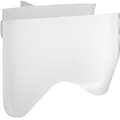
In 2010, the INBONE prosthesis was revised as INBONE II to help address some of the early failures. This iteration was an improvement in several ways. It provided a longer anterior-posterior length for the tibial component, added two anterior pegs to the talar component, and changed the saddle-shaped talus to a V-shaped sulcus design with increased stability[15] (Figure 1). A review of 59 INBONE I and II prostheses showed that the combined survival rate was an estimated 96.6% at a follow-up of 2 years. There were 5 revisions (8%) in INBONE I prostheses (4/5) and INBONE II prostheses, all for talar subsidence.[16] A more recent study found that the reoperation rates for 193 INBONE I and 56 INBONE II ankles were 18.5% and 15.9%, respectively, with failure rates of 6.0% and 2.6%, respectively, at 2 years postoperatively.[17]
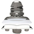
Photograph of the INBONE II ankle replacement. (Image courtesy of WrightMedical Group, Memphis, TN.)
The INBONE II is still used for both primary and revision ankle replacements. It is typically used for more severe deformities, however, such as a flat talus or major tibial bone loss, although some surgeons reserve it for revision only. Using this implant as a revision system is technically challenging, as demonstrated by the 31.4% complication rate in 35 Agility (DePuy Synthes) to INBONE revisions.[2]
Future reporting of midterm data and results on the INBONE II is expected to aid surgeons in making informed decisions about the system.
Recognizing the need for a less-invasive ankle without the use of a leg holder, Wright Medical developed a new prosthesis in 2013—the INFINITY Total Ankle System. The tibial component was modified to have ingrowth capability on three sides of the rectangular implant, with three spikes to impact into the cut tibial surface. The longer-stemmed tibia is still available for complex or revision cases. The talar component has two anterior spikes and an anterior and posterior chamfer with open sides, so it is possible to see under the prosthesis all the way across with fluoroscopy (Figure 2). The INBONE II talar component can also be used with the INFINITY tibial component in patients with a flat-topped talus, for which the chamfers would take out too much bone from the talar neck. The implant is too new on the market for outcomes data.

Figure 2.
Photograph of the INFINITY ankle replacement. (Image courtesy of Wright Medical Group, Memphis, TN.)
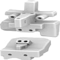
The Salto Talaris and Salto Talaris XT implants were acquired by Integra in October 2015 with the option to purchase the worldwide rights in 2017. The Salto Talaris fixed-bearing prosthesis has been available in the United States since 2006. This prosthesis was designed on the basis of its mobile-bearing counterpart in Europe—the Salto Total Ankle—first used in 1997. The fixed-bearing design came about after a radiographic study showed no anterior-to-posterior motion between the inferior surface of the tibial component and the superior surface of the polyethylene, showing that the implant was not functioning as a mobile-bearing system.[18]
These cobalt-chromium implants are single-coated with 200 μm plasma-sprayed titanium. The tibial component uses a central keel for fixation. During implantation, the trial tibial component is allowed to rotate to find the proper axis of rotation. However, the component is usually wedged between the malleoli, decreasing its rotation and undermining its ability to find its "home." The talar component has a conical shape with two different radii of curvature to match the morphology of the talus (ie, medial smaller than lateral). Although the lateral facet of the talus is resurfaced, the medial facet is not. The talar component also has a sagittal curved groove that, in theory, forces the foot into external rotation with dorsiflexion and allows internal rotation during plantar flexion. Four degrees of rotation around the center of talar curvature allows motion of the subtalar joint. A central peg is also present for stabilization.
The Salto Talaris XT consists of a flat cut talar component and a range of thicker polyethylene inserts aimed at revision and more complex primary applications (Figure 3). The XT implant uses the same articulation as the standard talar component, thus making it compatible with all Salto Talaris tibial and talar components.
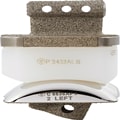
Photograph of the Salto Talaris XT with thickened polyethylene and flat cut talar component. (Image courtesy of Integra LifeSciences, Plainsboro, NJ.)
In a systematic review of 212 Salto Talaris ankles with a weighted follow-up of 34.9 months, only five prostheses (2.4%) needed revision (ie, three metallic component exchanges and two ankle arthrodeses).[19] There was no significant difference in revision rates between the design team/consultants and independent groups. The revisions were attributed to aseptic implant loosening, talar subsidence, tibial component subsidence, talar osteonecrosis, and tibial component aseptic loosening.
In a study of 300 patients who underwent 321 TAAs with Salto Talaris prostheses, 83.8% of patients experienced very good to excellent pain relief and 77.9% reported improved function at a mean follow-up of 38.9 months.[20] Interestingly, patients demonstrated improvement between postoperative months 12 and 24. At a mean follow-up of 20.1 months, eight patients required revision arthrodesis and two required revision TAA (2.3% and 0.6%, respectively).
A recent study of 78 patients who underwent 81 TAAs demonstrated a 97.5% survival rate at a mean follow-up of 5.2 years.[21] Seventeen patients (21.8%) underwent additional procedures after the arthroplasty, most commonly gutter débridement. Of those who had >2-year radiographic follow-up, 31% displayed evidence of lucency around either the tibial or the talar component.
The STAR was designed by Hakon Kofoed, MD, in collaboration with LINK AG, a German orthopaedic implant manufacturer, in 1978. The system was acquired by Small Bone Innovations (SBI) in 2009 and, ultimately, by Stryker in 2014. In total, five different versions of the STAR ankle have been implanted worldwide since 1981. Only the fourth-generation prosthesis has ever been available in the US market. Unlike the previous generations, the fourth-generation system is distinguished by the addition of a rough titanium plasma spray as a base coat to improve ongrowth. This design was used in the prospective clinical trials conducted in the United States under the investigational device exemption 8-year trial and ultimately received approval for widespread use in the country in 2009.
All the surgical instruments used in this system have been completely redesigned under the guidance of surgeons who participated in the clinical studies under the investigational device exemption. The goal in redesigning the instruments was to increase accuracy and reproducibility by changing the cuts from open blocks to captured cutting guides.
The STAR ankle is the only three-piece mobile-bearing design approved and available in the United States, although many mobile-bearing devices are available worldwide (Figure 4). The tibial tray has two 6.5-mm cylindrical bars for ongrowth, which allows for only 5 mm of distal tibial resection. The distal face of the tibial component is a flat, polished surface to allow unconstrained movement of the polyethylene component. The tibial component is wider anteriorly than posteriorly to mimic native anatomy. The talar component is symmetrically cylindrical, with medial and lateral wings to support the medial and lateral facets of the talus. A crest on the talar dome corresponds to a groove in the polyethylene component. The polyethylene insert has a meniscus that is congruent with both metal components. A recent study showed that there was preserved axial and sagittal motion at the tibial-polyethylene interface at a minimum follow-up of 1 year postoperatively.[22]
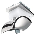
Photograph of the Scandinavian Total Ankle Replacement. (Image courtesy of Stryker, Kalamazoo, MI.)
To date, the fourth-generation STAR ankle has the longest follow-up data in the United States. In 2011, Mann et al[23] reported 91% metal component survivorship at 9.1-year follow-up. In 2015, Jastifer and Coughlin[24] reported 94.4% metal survivorship at a follow-up of 10.2 to 14.6 years. In 2012, Nunley et al[25] demonstrated 93.9% total implant survivorship rate at a mean of 60.1 months postoperatively.
The earlier generations of the STAR system have different prosthetic coatings. From 1989 through 1999, the prosthesis had a brushite and hydroxyapatite coating over smooth cobalt-chromium. In 1999, the coating was changed to a titanium plasma spray with a top layer of calcium phosphate, referred to as a double coat. These versions are available in Europe and are included in the European registry data but have never been available for use in the United States. Results with these prostheses are inferior to the results with the fourth-generation prosthesis in the United States. In 2013, Brunner et al[26] published results from the European registry with the first-generation STAR system, showing 70.7% survivorship at 10 years and 45.6% at 14 years. In 2016, Kerkhoff et al[27] showed a 10-year survivorship rate of 78% with the second-generation STAR system.
The Trabecular Metal Total Ankle is a fixed-bearing prosthesis with characteristics that differentiate it from other TAA devices. One notable feature of the system is that it uses a lateral transfibular approach, which requires a fibular osteotomy and transection of the anterior talofibular ligament, both of which necessitate postoperative repair.
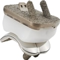
The aim of the transfibular approach is to maintain the integrity of the blood supply to the skin and to spare the deltoid ligament and minimize wound healing complications. In addition, the implant is designed to anatomically mimic the natural curvature of the tibia and talus. This results in less bone removal and more surface area contact. It also optimizes the perpendicular orientation between the prosthesis and the underlying bony trabeculae, which in turn resists subsidence (Figure 5).

Photograph of the Trabecular Metal Total Ankle. (Image courtesy of Zimmer Biomet, Warsaw, IN.)
The system relies on an external alignment system to hold the tibial plafond and talus in place while a burr removes articular cartilage and subchondral bone. The external alignment system also can be used for deformity correction.
The system is available in six sizes. The talar component is convex and is a cobalt-chromium-molybdenum alloy with a trabecular metal surface and a thin interlayer of titanium. It is available in separate right and left configurations. Its bicondylar articular geometry has a larger sagittal radius of curvature laterally than medially. The distal surface includes two fixation rails to facilitate stability. The articulating surface is on an 8° conical axis to replicate the geometry of the ankle. The tibial component is concave and symmetric. The tibial base is made from a Ti-6AL-4V alloy diffusion bonded to a trabecular metal surface. The proximal surface includes two fixation rails that are oriented perpendicular to the flexion-extension axis in the coronal plane to facilitate stability. The articular geometry of the implants is semiconforming in both the sagittal and coronal planes to allow semiconstrained motion. The polyethylene insert is highly cross-linked to achieve lower wear than conventional polyethylene.
In a study of 20 TAAs with a mean follow-up of 18 months, no fibular nonunion or delayed union was observed with the transfibular approach.[28]No implant failure was seen at 12 months postoperatively.
The Vantage Total Ankle System (Exactech) is a new prosthesis that was approved by the FDA in 2016. The ankle, which has a two-piece fixed-bearing design, is inserted through a standard anterior approach. It has four tibial and five talar sizes, each with right and left orientations (Figure 6). The polyethylene comes in thicknesses of 6 mm to 12 mm.

Photograph of the Vantage ankle replacement. (Image courtesy of Exactech, Gainesville, FL.)
The design is unique in that it was created from CT scans of normal and arthritic ankles[29] to provide maximum coverage in the anterior-posterior direction of the standard cut surface of the tibia. It has a fibular notch region that allows increased coverage without impinging on the fibula. It has a fenestrated cage design on the superior aspect of the tibial component, along with three spikes to help ensure bony ingrowth. The talar component has a rounding device that allows the surgeon to match the surface area of the talus to the implant rather than set the talar component on a flat surface. An anterior flange also helps prevent subsidence. The polyethylene component is inserted and fixed in place with a locking clip to allow easier removal and replacement in revision cases.
The design rationale for the Cadence Total Ankle System (Integra LifeSciences) was to develop an ankle replacement device that maintains anatomic kinematics while also offering the surgeon a multitude of fit options. The system is a cobalt-chromium alloy with a porous titanium plasma spray coating. The tibial component has two pegs and a posterior fin for fixation that is not prepped before insertion, allowing a solid press fit. The tibial component is available in standard and extra-long sizes, as well as left- and right-sided options to accommodate the patient's anatomy (Figure 7).
Photograph of the Cadence Total Ankle System. (Image courtesy of Integra LifeSciences, Plainsboro, NJ.)
Similar to the Trabecular Metal Total Ankle, the Cadence provides complete coverage of the resected tibia from the medial malleolus to the lateral edge of the tibia, thus necessitating left and right options. The talar component requires minimal bone resection and has two pegs for fixation. Articulation of the talar component is based on an 8° conical axis to replicate the natural kinematics of the ankle, and it has a sulcus design for rotational stability.
The polyethylene insert is made of highly cross-linked, ultra-high[FIGURE DASH]molecular-weight materials. The inserts can be anterior or posterior biased to help maintain the reduction of the talus under the tibial axis in cases of anterior or posterior subluxation (Figure 8).
Photograph of the anteriorly biased polyethylene of the Cadence Total Ankle System. (Image courtesy of Integra LifeSciences, Plainsboro, NJ.)
The INFINITY ankle replacement is unique in that it comes with an optional patient-specific guide technique called the Prophecy. A protocoled non[FIGURE DASH]weight-bearing CT scan of the patient's lower extremity is obtained, including views of the knee and ankle. The engineers then create a three-dimensional computer schematic that is approved by the surgeon and used to create patient-specific three-dimensional printed molds (Figure 9).
Photograph of the Prophecy patientspecific instrumentation. (Image courtesy of Wright Medical Group, Memphis, TN.)
The molds are placed on the patient's tibia and talus intraoperatively, and after radiographic verification that the alignment is correct, pins are placed in the bone through the molds. After that, the molds are removed and replaced with cutting guides. This technique has been 100% predictive within 3° of the preoperative alignment, 92% predictive of the tibial component size, and 36% predictive of the size of the talus.[30] The aberrancy in the talar sizing is likely a result of manual removal of bone from the gutters, which necessitates a narrower talar component. In addition, the measurements and guides are based on a non[FIGURE DASH]weight-bearing CT scan. Increased availability of weight-bearing CT scans may improve the accuracy of patient-specific instrumentation. The Prophecy is also available for the INBONE ankle replacement.
Fourth-generation and emerging ankle replacement designs share many factors: decreased distal tibial and talar bone resection, minimized disruption of the anterior tibial cortex, anatomically contoured distal tibial trays, and talar components with different curvatures of radii. Surgeons must balance the use of promising new designs with use of implants on which mid- to long-term outcome data are available. Informed conversations with patients are necessary to ensure that expectations are set and concerns are addressed in this burgeoning field. Surgeons need more complete and thorough data to help patients make informed decisions. Joint registries, research from high-volume institutions, and collaboration between institutions will be needed in the future.