Septic arthritis of the wrist is an uncommon condition, but one that can result in substantial morbidity. Timely identification and treatment is critical to patient care. No serum laboratory values have been shown to consistently confirm wrist joint infection. Thus, diagnosis is made based mainly on a thorough patient history, physical examination, and joint aspiration. When infection is suspected, aspiration of the wrist should be performed to confirm the diagnosis. Broad-spectrum antibiotics and joint aspiration or surgery are required to manage the infection and prevent sequelae.
Septic arthritis of the wrist is a rare but serious clinical entity that is typically defined by an infection within the radiocarpal joint; however, it can also include infections in the midcarpal and distal radioulnar joints.[1] In patients with severe infection or delayed presentation, the infection may extend into the carpal tunnel or deep soft tissues after exiting the wrist joint[1,2] (Figure 1). Thus, symptoms may vary depending on the location and timing of presentation. Although this joint-threatening condition has been described for nearly a century, few prospective trials are available to guide diagnosis and treatment because of the relative rarity of true septic arthritis in the wrist.
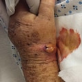
Clinical photograph demonstrating a case of a neglected septic arthritis of the wrist with extension of the infection out of the joint and into the snuffbox region of the wrist, resulting in a sinus that was initially assumed to be a superficial abscess.
Joint infection typically occurs because of direct inoculation by puncture or trauma, hematogenous spread from another site of infection, or contiguous spread from adjacent tissue.[3,4] After the organism is seeded, a reactive inflammatory process triggers the release of both cytokines and proteases, which ultimately cause cartilage destruction.[4]
For any infected joint left untreated, serious negative sequelae such as cartilage destruction and chondrolysis may begin as early as 8 hours after infection and can result in permanent joint dysfunction[5,6] (Figure 2). Further consequences, such as contiguous spread to the subchondral bone with resultant osteomyelitis, sinus formation, and even systemic sepsis, can result in cases of neglected septic arthritis.[7]
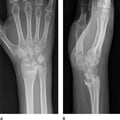
AP (A) and lateral (B) radiographs of the wrist demonstrating substantial radiocarpal destruction and chondrolysis from prolonged joint space infection.
Septic arthritis of the wrist often presents with a warm, erythematous, painful wrist without antecedent trauma[3] (Figure 3). Other conditions that mimic this presentation in the differential diagnosis include crystalline arthropathy, tenosynovitis, subcutaneous abscess, osteoarthritis, and cellulitis. Septic arthritis of the wrist is the most serious joint-threatening condition in the differential diagnosis.[8,9]

Clinical photograph demonstrating typical presentation of septic arthritis of the wrist, including a swollen, warm, erythematous, and painful wrist without antecedent trauma.
The true incidence of septic arthritis of the wrist is not well established.[9,10]In limited series, Mehta et al[10] reported that 23% of upper extremity joint infections occur in the wrist, although Skeete et al[9] reported only a 5% overall incidence. Similarly, a large study by Yap and Tay[11] confirmed only 40 cases of septic arthritis of the wrist over an 11-year period. Thus, although the incidence of septic arthritis of the wrist is not established, the diagnosis can be assumed to be uncommon. However, because of the high morbidity of permanent wrist dysfunction, vigilant diagnosis and treatment is indicated in patients with suspected wrist joint infection.
A thorough history and physical examination remain critical components of the evaluation of a suspected wrist infection in a painful wrist without antecedent trauma.[12] Wrist pain, stiffness, diminished hand and wrist strength, fever, and chills, along with a history of recent illness or distant infection, direct joint inoculation, or recent surgery are common issues.[11,12]Among these issues, pain and stiffness are the most common, and as few as 18% of patients may present with a true fever.[11] Septic arthritis of the wrist most commonly affects adults in the fifth through seventh decades of life, although the condition can affect persons of almost any age.[1,9–11,13]
Comorbidities such as chronic kidney disease, diabetes, alcoholism, active intravenous drug use, and any condition or medication resulting in an immunocompromised status should increase suspicion for septic arthritis.[11,13] Yap and Tay[11] noted that >82% of wrist joint infections identified in their study were found in patients considered to be immunocompromised. Similarly, patients who are undergoing chronic steroid therapy or who take disease-modifying antirheumatic drugs may be at a higher risk compared with the general population.[13]
Patient age may influence the diagnosis: disseminated gonorrhea, chlamydia, syphilis, Reiter disease, and Lyme disease are all more common in young, otherwise healthy patients.[14] In cases in which septic arthritis of the wrist is the result of disseminated sexually transmitted disease, a history of recent unprotected sex, related symptoms such as burning with urination, or involvement of other joints may be elicited.[15–17] Although monoarticular arthritis in large joints, such as the knee or hip, are more common presentations of Lyme arthritis, this diagnosis must be considered, particularly in endemic regions.[17–19]
Similarly, in older patients, conditions such as gout, pseudogout, and cellulitis are most commonly mistaken for a septic wrist joint. Skeete et al[9]identified these other conditions as causing a hot, painful, atraumatic wrist in 67% of 104 patients in a retrospective cohort, but only 5 patients had a septic wrist joint. True septic arthritis was found in only 12 of 52 patients in a 10-year review by Mehta et al[10] and in only 40 patients in an 11-year review by Yap and Tay.[11] Yap and Tay[11] did not indicate the total number of patients treated in that period.
On physical examination, patients with septic arthritis of the wrist typically have a warm, swollen, and erythematous wrist joint. Other findings include pain and guarding on passive wrist range of motion and diminished active total arc of motion. However, other common conditions can present similarly, including crystalline arthropathy, tenosynovitis, and cellulitis. Therefore, objective laboratory markers in serum and joint fluid can help make the diagnosis.[9]
Serum analysis in the setting of any suspected infection includes blood cultures, a complete blood count (CBC), erythrocyte sedimentation rate (ESR), and C-reactive protein (CRP) level. Elevation of these serum markers indicates only a nonspecific inflammatory process rather than a specific diagnosis for septic arthritis of the wrist.[9–11] In a study by Mehta et al[10] of 52 patients with upper extremity septic arthritis, only 58% had an elevated white blood cell (WBC) count, which corroborated results of previous studies.[20] Similarly, Skeete et al[9] examined a series of septic wrist cases and were unable to distinguish those with a wrist infection from other diagnoses, such as crystalline arthropathy, cellulitis, tenosynovitis, or cellulitis on the basis of serum markers such as ESR or CRP level.
Because of the broad differential diagnosis and limited diagnostic value of serum laboratory markers, joint aspiration is often used to help diagnose septic arthritis of the wrist. For this procedure, an 18-gauge needle on a 3- to 5-mL syringe is inserted into the wrist joint dorsally, just distal to the Lister tubercle (Figure 4). Because the wrist is a small joint and typically yields only a few milliliters, even in cases of infection, so-called dry taps or an arthrocentesis without enough fluid to perform a full analysis are common, potentially contributing to a high false-negative rate.[9] Furthermore, unlike with larger joints such as the hip and knee, no specific laboratory values on joint fluid analysis are pathognomonic for septic arthritis of the wrist. Although a synovial fluid WBC count >50,000 cells/mm3 strongly suggests an infection, other conditions may also present with a similarly high count.[12,21] Similarly, no diagnostic neutrophil percentage has been established to be pathognomonic for septic arthritis of the wrist; therefore, results must be interpreted cautiously.
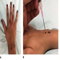
Clinical photographs demonstrating needle aspiration of the radiocarpal joint with an 18-gauge needle inserted into the dorsal wrist capsule. A, Planned needle placement (black circle) just distal to the Lister tubercle (open circle). B, The needle is oriented parallel to the joint surface of the distal radius with a slight distal to proximal angulation.
Analysis of joint fluid aspirate for crystals may establish the diagnosis of gout or pseudogout, depending on their shape and birefringence. Crystalline arthropathy frequently affects the wrist joint and can often present with signs and symptoms that are difficult to distinguish from a true wrist infection.[22]Several cases of concomitant crystalline and septic wrist pathology have been reported; therefore, infection still must be ruled out even after identification of synovial crystals.[23–25]
Perhaps most important, a positive Gram stain and culture can both confirm the diagnosis of septic arthritis and guide antibiotic therapy. Although many organisms may be responsible for wrist joint infection, Staphylococcus aureus remains the most common.[9,11,12,26] In up to 40% of cases of septic arthritis of the wrist, no organism is identified; this knowledge, combined with the frequency of arthrocentesis with insufficient fluid for analysis, illustrates the importance of the patient history and physical examination in establishing a diagnosis.[1,10,12,27] Therefore, in the typical situation for which limited joint fluid is available for analysis, priority should be given to cultures, Gram stain, and crystalline analysis over cell count. In regions endemic for Lyme disease or for cases in which Lyme infection of the wrist is suspected, culture is almost always negative and polymerase chain reaction (PCR) testing of the synovial fluid may be added, although results can take several days.[18] Similarly, gonococcal arthritis is notoriously difficult to culture, and PCR may be added in these cases as well.[28]
Along with history, physical examination, and laboratory workup, radiographs of the wrist also should be obtained. In cases of long-standing or delayed presentation of a septic wrist, radiographic findings of symmetric joint destruction and chondrolysis may be present (Figure 2). In addition, the presence of hardware in the wrist should increase concern for a septic process, as should any foreign body in the joint.[12] Conversely, the presence of any fracture, dislocation, inflammatory arthritis, or osteoarthritis may provide alternate explanations for wrist pain and swelling. MRI may be most useful in identifying collections or edema elsewhere in the hand or wrist. MRI also distinguishes involvement of the infection in locations beyond the radiocarpal joint, such as the midcarpal, carpometacarpal, or distal radioulnar joints. Although both MRI and ultrasonography can identify pathologic fluid collections or joint effusions, findings may be analogous to cases of inflammatory arthritis or crystalline arthropathy and therefore are not diagnostic.[12,29]
Management of septic arthritis of the wrist includes isolation of the organism and joint aspiration or surgical drainage combined with antibiotics and temporary immobilization of the joint.[3,11,12,27] Delayed treatment can result in substantial morbidity; therefore, early aspiration and/or surgical drainage is recommended.[27] Good to excellent results have been reported when surgery is performed within 10 hours after diagnosis, with outcomes worsening when treatment is delayed beyond 16 hours.[3] No surgical intervention is warranted for patients with Lyme or sexually transmitted disease or reactive arthritis because disease-specific antibiotics are the mainstay of treatment in these cases.[19,28,30,31]
The standard surgical management of septic arthritis of the wrist involves open arthrotomy of the radiocarpal joint, with extension into the midcarpal space as needed.[3,12,32] The radiocarpal joint typically is accessed using a standard dorsal approach, which begins with a midline incision dorsally, followed by arthrotomy of the wrist joint between the third and fourth extensor compartments, although several other techniques have been described[3,33–35] (Figure 5). At the time of wrist joint arthrotomy, aerobic and anaerobic cultures should be obtained. Although an immunocompromised state or an abnormal presentation should increase concern for atypical infection, acquiring cultures for both tuberculosis and fungal infections is recommended in nearly all cases.[26,32] After cultures are obtained, the radiocarpal and midcarpal joints should be irrigated with normal saline. If clinical concern warrants it, exposure and washout of the distal radioulnar joint can also be performed.[26,27] A volar approach to the radiocarpal joint is uncommonly indicated, but a volar approach may be used for patients in whom long-standing disease has resulted in concomitant deep-space infections of the volar hand or carpal tunnel.
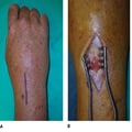
Photographs showing the dorsal approach to the radiocarpal joint, with the planned incision centered over the wrist (A), ulnar to the Lister tubercle (circle). B, The joint is arthrotomized between the third and fourth extensor compartments, where cultures are obtained before a formal joint irrigation.
Arthroscopic management of wrist septic arthritis can also be effective. Potential additional benefits of arthroscopy include smaller incisions, better joint visualization, fewer procedures, and shorter hospital stays.[12,26,27]Typically, the 3–4 portal is used to visualize the radiocarpal joint with a 2.4-mm diameter arthroscope. Joint lavage is delivered via the arthroscope. The 6R or 6U portal is used to insert a shaver to manage loculations and synovitis, as well as for joint fluid suction.[27,36] An additional midcarpal and/or distal radioulnar portal can be placed as needed. Because the distal radioulnar portal is smaller, a 1.9-mm diameter arthroscope may be required.[37,38]
No evidence has demonstrated superiority of open or arthroscopic drainage compared with simple needle aspiration of the wrist[1,39,40] (Figure 4). However, needle aspiration is most commonly used in patients in whom surgical intervention is medically contraindicated. Although serial needle aspiration may obviate the need for surgical intervention, several aspirations may be required until symptoms or the effusion resolve.[1] Moreover, needle aspiration is insufficient in patients with septic arthritis of the wrist resulting from infected hardware or any foreign body that warrants surgical removal.[1]
Antibiotics should be withheld until after cultures have been obtained, after which an empiric broad-spectrum coverage agent is appropriate.[1]Recommended empiric intravenous treatment includes vancomycin plus another antibiotic that treats methicillin-resistant S aureus (MRSA), aerobic and anaerobic bacteria such as ceftriaxone, or ampicillin-sulbactam. Ideally, the Gram stain findings should direct antibiotic treatment. S aureus is the most frequently identified organism of many species identified in cases of septic arthritis of the wrist.[10,11,13,26] Streptococcal species are the second most common, followed by gram-negative organisms such as Pseudomonasand mixed-flora infections.[11,13] Although methicillin-sensitive S aureus is most commonly identified, evidence exists of increased prevalence of community-acquired MRSA infections.[41,42] Therefore, in areas with a high prevalence of MRSA infections, empiric treatment with vancomycin, clindamycin, trimethoprim-sulfamethoxazole, or daptomycin should be considered.[1,41,42]
Final laboratory cultures should be used to determine the antibiotic used. Typically, intravenous treatment is indicated for 1 to 2 weeks, followed by oral treatment for another 2 to 4 weeks.[13,43,44] However, the optimal duration of intravenous and oral antibiotics has yet to be determined and should be based on the clinical overview, including the severity of the infection, the patient's overall health, and the organism identified. Unsuccessful treatment is uncommon with appropriate antibiotics and joint drainage, and can often be attributed to delays in diagnosis or an atypical organism (eg, a mycobacterium).[13,45]
Outcomes for the management of septic arthritis of the wrist are generally favorable when treatment is timely, using either aspiration or surgery and concomitant antibiotic therapy. In a series of 29 wrists, Rashkoff et al[3]showed the best results when arthrotomy was performed within 10 hours of diagnosis. Overall, no recurrent infections occurred at follow-up (range, 6 months to 9 years). In a cohort of 40 patients, Yap and Tay[11] reported a 6-month mortality rate of 15% with a mean time of 6.8 days from presentation to surgery.
No large prospective studies exist on optimal treatment. However, several authors have retrospectively examined outcomes for wrist joint infections. Goldenberg et al[40] examined a series of 59 joint infections, 7 of which involved the wrist, and all of which were treated with needle aspiration rather than surgery. All seven patients reported good outcomes at a minimum follow-up of 3 months. Sammer and Shin[12] compared 19 wrists treated with open irrigation and débridement with 21 wrists treated arthroscopically in patients with primary septic arthritis of the wrist. Although most patients in both groups required at least one additional procedure, no substantial difference was noted between groups with regard to the number of repeat surgeries or length of hospital stay. However, no data are available regarding postoperative clinical outcomes.
Although septic arthritis of the wrist is uncommon, it can result in substantial morbidity and mortality if it is not recognized early.[1,8,9,11,45] Urgent evaluation with a history and physical examination should help the treating physician determine the diagnosis, although blood cultures and serum and joint fluid analysis often complete the clinical picture.[1,9] No specific laboratory values have been identified as pathognomonic for septic arthritis of the wrist.[9–12] In this context, when limited joint fluid is available for laboratory evaluation, priority should be given to Gram stain, culture, and crystal analysis results.
Management includes needle aspiration or surgical drainage allowing empiric intravenous antibiotic therapy.[11,12,26,27] Typically, a single débridement is successful; however, patients with substantial illness or comorbidities may require multiple procedures.[45] Although S aureus is the most commonly isolated organism in cases of wrist septic arthritis, broad-spectrum antibiotics should be initiated until cultures identify the causative organism.[10,11,13,26] Missed diagnoses can result in permanent sequelae, including cartilage and joint destruction, contiguous spread to nearby tissues, and even bacteremia or sepsis.[5–7] To date, no large prospective studies have elucidated an optimal surgical treatment, although with timely débridement and appropriate antibiotic coverage, favorable outcomes can be expected in most patients with septic arthritis of the wrist.[6,11–13]