We aim to critically review the effectiveness and safety of coccygectomy with special regard to long-term outcomes.
Coccygectomy was performed in our clinic in 38 patients between 1990 and 2019. All these patients (32 females vs. 6 males) have failed to respond to conservative treatment for at least 6 months prior to surgery. All patients were available for follow-up after mean 12,3 years (2 months to 29 years, 11 patients had a minimum FUP of 24 years). We evaluated all patients clinically and radiologically.
Nineteen patients reported traumatic and 17 patients reported idiopathic onset of their symptoms; one patient had clinical symptoms after childbirth and another patient had coccygodynia after extensive low back surgery. 36 of our 38 patients were free of pain at least 6 months after surgery and had good or excellent clinical results according to the VAS which improved from 6.37 (SD 1.08) preoperatively to 0.68 (SD 0.99) at the recent follow-up. Two patients showed an ODI > 22 at the recent follow-up (24 and 28) and 32 had an ODI equal or under 4. There was no statistical significant difference in terms of clinical outcome between the different radiological types of the coccyx. Postoperative complications were rare: 1 superficial infection and one re-operation 6 months after initial surgery due to an pre-existing exostosis which had not been removed at the index surgery; no neurological complications and no major bleeding occurred. No patient had recurrent onset of coccygodynia. 37 out of 38 patients would have coccygectomy again.
Coccygectomy is a safe treatment option in patients with coccygodynia and shows excellent long-term results. We recommend to perform coccygectomy if patients fail to respond to conservative treatment for 6 months.
IV
The definition of coccygodynia is discomfort or pain in the lowest part of the spine, the coccyx. The condition was first defined by Simpson in 1958 [1] and is mainly triggered by direct trauma [2, 3]; indirect injuries; and the summation of micro-injuries [4] or by abnormal mobility of the tailbone. If the etiology of coccygodynia is not post-traumatic or mobility related it is called idiopathic. Organic reasons like neurogenic pain [5], arachnoid cysts, pilonidal sinus, and neoplasms [3], etc., have been described and have to be considered as possible causes of pain during the diagnostic algotythm [1, 3,4,5,6]. Sacrococcygeal pain is five times more likely in women than in men [5, 6]. It is widely accepted that the therapeutic algorithm is as follows [7,8,9,10,11,12,13,14,15].
The first line of treatment consists of conservative treatment options such as non-steroidal anti-inflammatory drugs, sitting aids, physiotherapy, manual treatments such as joint mobilisation, or the repetitive injection of corticosteroids. Up to 85% of patients respond to these treatments and heal [9]. If the remaining patients do not respond to these treatment protocols, coccygectomy has to be considered as the next therapeutical option.
Despite favourable clinical data [7, 9, 11, 12] surgical excision of the coccyx for the treatment of therapy-resistant coccygodynia is often delayed for a long time due to various concerns. The main reasons are well reported high infection rates [10, 12,13,14], the rarity of the condition [12], and absent ill-defined selection criteria to identify patients who may benefit from this type of surgery.
It is not clear yet if there is a difference in terms of long-term outcome between different aetiologies in patients treated with coccygectomy and only few studies concerning the long-term follow-up after coccygectomy exist. Many patients suffer from coccygodynia over years until they are treated surgically and adequately.
The purpose of this retrospective study is to report on long-term outcome and the effectiveness of pain relieve and safety of coccygectomy with a critical review of the current literature in order to provide a decision aid when to perform coccygectomy.
In our university clinic, coccygectomy was performed on 38 patients between 1990 and 2019. Coccygodynia was diagnosed when a history of pain in the sacrococcygeal area aggravated by sitting and re-created by direct external examination of pain confined to the mobile coccyx was present. Our conservative treatment regime prior to surgery consists of at least 3 months physiotherapy, NSAIDs, repeated local steroid injections combined with anaesthetics, and digital manipulation. All these patients have failed to respond to conservative treatment for at least 6 months. All 38 patients (32 females vs. 6 male) were available for evaluation.
Recorded data includes the patient’s demographics, duration of symptoms, aetiology, medications, comorbidity including spinal pathology, treatment prior to surgery, functional status, and a radiologic evaluation. All patients completed a questionnaire, including the visual analogue scale pain indicator (VAS), the Oswestry Disability Index (ODI), and the Quebec Back Pain Disability Scale (QBPDS).
The VAS, an easy to use and reliable measure of pain, uses a range of 100 mm for assessing pain intensity, ranging from zero (free of pain) to 100 mm (very severe pain) [15]. An ‘‘excellent’’ outcome was achieved with complete absence of pain or significant improvement of pain; this was operationalized as a VAS less than 2 of 10 and an increase in quality of life. A significant improvement in pain and a VAS less than 3 out of 10 was classified as a ‘‘good’’ result. A moderate improvement of pain and a VAS less than 6 out of 10 was classified as a ‘‘fair” outcome. Unchanged pain symptoms, an increase in complaints, or a VAS at the time of the investigation of more than 6 out of 10 was classified as a ‘‘poor’’ result.
The ODI was developed to determine limitations of various activities of daily living; is widely used as condition-specific measure for spinal disorders; and has a proven record of reliability, validity, and responsivity. It is scored from zero (no disability) to 100% (complete disability), with a score of 22 or higher considered as significant ADL disability [16]. The threshold for successful treatment was based on an overall ODI score of < 22 points at the last follow-up [16, 17].
The QBPDS is a 20-item self-administered instrument designed to assess the level of functional disability in individuals with back pain [18].
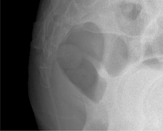
For radiological assessment, we were using both the classification system according to Postacchini and Massobrio [15], and the Maigne classification [19]. In the Postacchini classification: type I means a slightly forward curved coccyx; type II is more markedly curved, with the coccyx pointing straight forward; type III is sharply angled anteriorly; and type IV shows subluxation of the sacrococcygeal or intercoccygeal joints (Fig. 1).
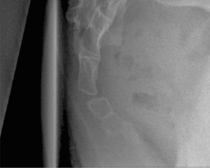
Maigne describes 3 types of coccyges: type I shows a forward curvature more than 25°, type II is a displaced or posteriorly subluxed coccyx, and type III tailbone is immobile with spiculae. Spiculae are defined as morphological abnormality in the form of a small bony excrescence on the dorsal aspect of the tip of the coccyx [20]. The sacrococcygeal angle was evaluated in preoperative lateral view radiographs (Fig. 2).
Our operative technique is as follows: all patients underwent total or partial coccygectomy by subperiosteal dissection as originally described by Key [21] through a longitudinal midline incision while in a modified prone position. The proximal bone ends were chamfered. Closure was performed by reapproximating the fascia and closing the subcutaneous layer and the skin. All patients had a drain for one day. Single-shot Cefazoline was used perioperatively as antibiotic prophylaxis. No bowel preparation was used preoperatively. Sutures were removed two weeks following surgery and patients were allowed to mobilise and sit as tolerated.
Descriptive statistics were calculated for all patient characteristic variables. The VAS, ODI, and QBPDS scores were calculated for each patient. Statistical significance was set at p = < 0.05.
Statistics were performed using Fisher exact and Chi-square analysis for categorical variables. The Student’s t-test was used to analyse normally distributed continuous variables, and the Mann–Whitney test for non-normally distributed, noncontinuous variables.
In our 38 patients the mean age at surgery was 36.7 years (13.4–67.8, female 36.1 vs. male 39.6)) and the ratio female/male was 5.3:1 which is consistent with the literature [9]. Besides two patients with diabetes mellitus no relevant comorbidities were found. Follow-up was performed after mean 12.3 years (2 months to 29 years) at a mean age of 49 years (17.9–82). Eleven patients had a minimum FUP of 24 years and 20 patients more than 10 years. Regarding at aetiology 19 patients report traumatic onset of their symptoms, one patient had clinical symptoms after childbirth and one patient reported onset of pain after many hours of cycling. In the remaining 17 patients, no pathognomonic causality was found, therefore coccygodynia was classified as idiopatic in these cases.
36 of our 38 patients were free of pain at least 6 months after surgery and had good or excellent clinical results according to the VAS, ODI, and QBPDS at the recent follow-up. The only patient reporting persistent pain was one with idiopathic coccygodynia and no trauma in history. Pain on the VAS improved from 6.37 (SD 1.08) preoperatively to 0.68 (SD 0.99) at the recent follow-up. Two patients showed an ODI > 22 at the recent follow-up (24 and 28) and 32 had an ODI equal or under 4.
Radiological assessment was performed in all patients. According to the Postacchini classification we found 16 type I, 14 Type II, 5 Type III, and 3 Type IV coccyges. Using the Maigne classification, there were 36 type I, 2 type II and no type III configuration. The mean number of coccygeal segments was 3,1 (2–4).
There was no statistical significant difference in terms of outcome between the different radiological types of the coccyx. Postacchini Type III patients seem to have more pain preoperatively and slightly less favourable outcomes postoperatively compared to Type I, II and IV (p 0,145).
We removed the coccyx always at the level of hypermobility or instability as assessed during surgery. We performed 9 total and 31 partial coccygectomies and found no difference in the outcome between both groups. There was no difference between the different aetiologies and the long-term outcome as well as there was no deterioration of outcome in the course of time.
Under perioperative antibiotic prophylaxis 1 superficial infection occurred. It healed with conservative treatment and oral antibiotics. All except one patient would have had surgery done again.
The painful tailbone is a rare condition with an incidence of under 1 percent of all low back pain patients [6, 12]. Because of its rarity many physicians are not aware of its aetiologies, diagnostics, and treatment strategies. For this reason, adequate diagnostics and sufficient therapy, especially surgery, is often delayed. We want to show that coccygectomy is a safe treatment option in the long term for patients suffering from post-traumatic and hypermobility related coccygodynia resistant to conservative treatment for 6 months.
Patients suffering from coccygodynia can be divided in two main groups following their aetiology: traumatic, and idiopathic coccygodynia [22].
Idiopathic coccygodynia has been described as the absence of any obvious pathologic changes involving the coccyx, although this is considered a diagnosis of exclusion. SHORTENED However, most cases are associated with abnormal mobility of the coccyx at the sacrococcygeal joint or within one of its segments which may trigger a chronic inflammatory process in the unstable section leading to degeneration of this structure. Conservative treatment strategies remain the first line of therapeutical options for coccygodynia consisting of NSAIDs or other analgesics, physical therapy, reduced or adjusted sitting, or joint mobilisation [24]. Repeated local administration of steroids and anaesthetic represent another non-surgical approach to therapy refractory pain. Wray et al. report 79% pain free patients after repeated injections. Additional digital manipulation enhanced this up to 89% [10]
In most patients undergoing surgery treatment consists either in (partial) excision of the mobile segment or total coccygectomy. With a success rate of 94% in the present study we are above the cumulative effectiveness of 84% Karadimas et al. [9] reported in their review. Coccygectomy in subjects with normal coccygeal mobility seem to have less favourable results [23]. Due to our selection criteria for coccygectomy all our patients showed abnormal mobility in sacrococcygeal joint or intracoccygeally. We did not see any statistical relevant relationship between traumatic and non-traumatic, respectively, different intercoccygeal angles with pain intensity or outcome.
Superficial and deep wound infection are the most frequent complications in coccygeal surgery and occur in up to 22% of the cases. The overall complication rate is about 10% [9]. Developments in intraoperative wound care and prophylactical intravenous administration of antibiotics have reduced these infections down to 0%, e.g. in the study of Doursounian [25]. With one superficial infection which could be treated conservatively, our infection rate was 2.6%. Surgical dissection of the coccyx according to Key [21], combined with perioperative antibiotics and a drain for one day, and no bowel preparation, seems to be safe and reproducible. The reason for the observed low infection rate could be that all but two coccygegtomies have been performed by one experienced senior surgeon in mainly young patients (76% under 50 years of age) with few comorbidities.
Clinical and radiological differences between idiopathic and traumatic coccygodynia seem to affect pain intensity preoperatively and clinical outcome postoperatively. In contrast to Kim et al. [22], we found no significant differences between the traumatic (T) and idiopathic (I) coccygodynia in terms of the pain score. They suggest the increased intercoccygeal angle as a possible cause of idiopathic coccygodynia [22].
Kim and Suk [22] found significant differences between the intercoccygeal angle of patients with traumatic (47,9°) and idiopathic Coccygodynia (72,2°). In our patients no significant difference in the intercoccygeal angle was found (trauma: 50,5°, idiopathic: 57,3°). With a female to male ratio of 5.3:1 our study population was similar to previous studies [9].
There are limitations to the study. This is a retrospective analysis with its inherent recall and information biases. As we focused on instability related causes for coccygodynia we did not include other causes to surgical treatment. We were not able to determine the incidence of coccygodynia in our clinical documentation system in the last 30 years. Baseline ODI and QBPDS values have not been evaluated preoperatively in our institution therefore we were not able to demonstrate the rate of improvement.
In our clinical setting, therapy resistant instability related coccygodynia was the trigger to indicate coccygectomy and we found good results—this is why we advise to focus on patients with clinically detectable instability to set for surgery.
The results of this retrospective long-term follow-up cohort study suggest that coccygectomy in traumatic and idiopathic hypermobility caused coccygodynia is a safe treatment option and shows excellent long-term results up to the third decade. Therefore, we are convinced that with a strict diagnostic algorithm, it is not necessary to delay surgical treatment for longer than 6 months.
Open access funding provided by Kepler Universitätsklinikum Linz.. There was no external funding source for this study.
Conceptualization: Rainer Hochgatterer; Methodology: Rainer Hochgatterer, Tobias Gotterbarm, Lorenz Pisecky; Formal analysis and investigation: Rainer Hochgatterer, Manuel Gahleitner, Jakob Allerstorfer, Jakob Maier; Writing—original draft preparation: Rainer Hochgatterer; Writing—review and editing: Lorenz Pisecky, Tobias Gotterbarm, Gerhard Großbötzl, Rainer Hochgatterer; Resources: Tobias Gotterbarm; Supervision: Lorenz Pisecky
All authors declare that they have no conflict of interest.
Ethical approval was waived by the local Ethics Committee of the Kepler University Clinics in view of the retrospective nature of the study and all the procedures being performed were part of the routine care.
Informed consent was obtained from all participants included in the study.
Patients signed informed consent regarding publishing their data.
Springer Nature remains neutral with regard to jurisdictional claims in published maps and institutional affiliations.
Open Access This article is licensed under a Creative Commons Attribution 4.0 International License, which permits use, sharing, adaptation, distribution and reproduction in any medium or format, as long as you give appropriate credit to the original author(s) and the source, provide a link to the Creative Commons licence, and indicate if changes were made. The images or other third party material in this article are included in the article’s Creative Commons licence, unless indicated otherwise in a credit line to the material. If material is not included in the article’s Creative Commons licence and your intended use is not permitted by statutory regulation or exceeds the permitted use, you will need to obtain permission directly from the copyright holder. To view a copy of this licence, visit http://creativecommons.org/licenses/by/4.0/.